The human foot, a marvel of biomechanics, propels us through life. But what happens when a silent enemy lurks within? Charcot foot, a deceptive condition often associated with diabetes, dismantles the intricate structure of the foot from the inside out.
This collection of Charcot foot images transcends the limitations of X-rays. We are showcasing not just the fractured bones, but the often-unseen consequences of Charcot foot. Witness the inflammation, the distorted architecture, and the story signs of a foot in distress.

Through Charcot foot images, you will gain a profound understanding of the devastating nature of Charcot foot. But more importantly, we illuminate the path towards early detection and intervention. That empowers individuals to safeguard their mobility and prevent the silent destruction of this deceptive condition.
What is a Charcot foot image?
Charcot foot is a condition that is often associated with diabetes and presents a unique challenge. While description can outline the symptoms and consequences, visual representations offer a powerful tool to truly understand the hidden devastation within the foot.

Charcot foot images encompass two main categories. Each reveals crucial details about this deception condition:
1. Clinical Photographs:
These images capture the physical changes associated with Charcot foot. Also serves as a visual confirmation of the condition’s progression. They may showcase:
Swelling and redness: Often the initial signs, of these changes can be localized or encompass the entire foot. That indicates inflammation and underlying damage.

Deformities: As the condition progresses, the foot can lose its normal shape. It can develop deformities like rocker-bottom feet or clawed toes, which can significantly impact movement and stability.

Skin breakdown and ulceration: In later stages, the weakened and deformed foot becomes susceptible to pressure points and skin breakdown. It leads to ulcer formation. These ulcers can become infected and pose a serious health risk.

2. Medical Scans:
These advanced imaging techniques provide detailed insights into the underlying bone and joint damage. These reveal the true extent of the destruction:
X-rays: These scans visualize bone structures. Often shows fractures and dislocations in various stages of healing or progression, disrupted joint alignment, and changes in bone architecture.

MRI scans: MRI scans offer superior soft tissue visualization. MRI scans are used maximum because they can detect joint, effusion, early-stage bone marrow edema, and microfractures. MRI scan is crucial for early diagnosis and intervention.

Charcot Foot Image
Charcot foot images go beyond undiluted optical statements; they pretend as a cool testament to the tricky nature of this condition. A broken bone with a clear fracture line, Charcot foot’s extermination uncover subtly.

Images capture the perverted landscape of the foot, where swelling changes into deformities. Skin breakdown also expresses the fragility beneath.

In terms of Charcot foot images, X-rays delve deeper and expose the dislocated and fractured bones. X-ray is a silent testament to the internal disorder shaped by this noiseless destroyer.

These images serve as a full reminder that the actual story of Charcot foot lies not only just in its symptoms but also in the hidden damage that lurks under the surface.

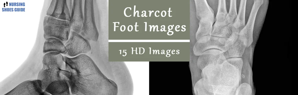
Here are 15 high-quality Charcot foot images that can help you easily understand how it looks and hurts on the foot. 4 stages clarify the whole Charcot joint foot:
Stage 0:

Stage 1:

Stage 2:

Stage 3:

Conclusion
Charcot Foot images nurses as a complete reminder that the true story of this condition lies not just in its symptoms, but in the silent eradication lurking under the surface. By appearing beyond the initial swelling and redness, we witness the deceptive progression of bone fractures, joint dislocations, and soft tissue erosion.
These visuals underscore the critical importance of early detection and intervention. Regular foot examinations, coupled with an awareness of the deceptive nature of Charcot foot, empower individuals to protect their mobility and prevent the weakening consequences of this condition.
While the visuals may reveal the wasting path of Charcot foot, they also enlighten the path to prevention, ensuring that individuals can maintain a healthy and active life.
Leave a Reply